Ultrasound improves image accuracy by producing real-time images, allowing clinicians to optimise image settings during the examination, and guiding procedures with continuous visual feedback. Newer technologies such as micro-ultrasound, Doppler imaging, elastography, MRI-ultrasound fusion, and AI-assisted image analysis are further improving the detection and targeting of clinically significant prostate cancer.
The question many clinicians and patients ask is: How does ultrasound improve image accuracy? Read this article to learn more about it.
On this Page
How Does Ultrasound Improve Image Accuracy?
In urology, ultrasound is especially important because it provides real-time guidance during prostate biopsy, allowing the clinician to continuously visualise the prostate, surrounding structures, and biopsy needle. This significantly improves procedural accuracy compared with performing biopsies without image guidance.
The answer lies in a combination of physics, equipment quality, operator expertise, and technological advances. Modern ultrasound systems can now produce remarkably detailed images, helping clinicians distinguish normal tissue from suspicious lesions more accurately than ever before. High-frequency probes, advanced beamforming, image processing algorithms, Doppler imaging, elastography, contrast-enhanced ultrasound, and artificial intelligence have all contributed to substantial improvements in diagnostic performance.
What Does Ultrasound Image Accuracy Mean?
Image accuracy refers to how faithfully an imaging system represents the anatomy being examined.
An accurate ultrasound image should clearly display:
- Prostate boundaries
- Internal gland architecture
- Suspicious lesions
- Blood vessels
- Needle position during biopsy
- Surrounding organs and tissues
Greater ultrasound image accuracy enables clinicians to identify abnormalities with more confidence while reducing the likelihood of missing clinically significant disease.
For prostate biopsy, improved image accuracy translates into:
- Better lesion localisation
- More precise biopsy targeting
- Reduced sampling error
- Improved diagnostic confidence
- Fewer repeat biopsy procedures
These benefits have become increasingly important as prostate cancer diagnosis has shifted from systematic random sampling towards targeted biopsy based on imaging findings.
How Ultrasound Creates Images?
Unlike CT scans or X-rays, ultrasound uses high-frequency sound waves rather than ionising radiation.
The ultrasound transducer sends sound waves into the body. As these waves encounter tissues of different densities, some are reflected to the probe while others continue deeper into the body. The machine measures:
- Time taken for echoes to return
- Strength of reflected echoes
- Direction of returning signals
Powerful image-processing software converts these returning echoes into a grayscale image displayed almost instantly. Because this process occurs many times per second, clinicians receive continuous real-time imaging rather than a single static picture. This allows them to adjust probe position immediately if visualisation is suboptimal.
Why Real-Time Imaging Improves Accuracy?
One of ultrasound’s greatest advantages is that it provides live imaging. Rather than relying on images captured earlier, clinicians can:
- Rotate the probe
- Adjust scanning angles
- Improve visualisation
- Follow moving anatomy
- Monitor needle placement continuously
This is particularly valuable during transperineal prostate biopsy. As the biopsy needle advances, its position can be monitored throughout the procedure, allowing adjustments before tissue samples are taken. Real-time visualisation reduces targeting errors and helps ensure tissue is collected from the intended region of the prostate.
Factors improve Ultrasound Image Accuracy
1. Higher Frequency Transducers
Image resolution largely depends on ultrasound frequency. Higher-frequency probes produce:
- Sharper images
- Better tissue differentiation
- Improved edge definition
- Greater detail of superficial structures
The trade-off is reduced depth penetration.
Conventional prostate ultrasound typically uses frequencies between 7 and 12 MHz. Newer micro-ultrasound systems operate at approximately 29 MHz, providing nearly three times the spatial resolution of conventional transrectal ultrasound. This higher resolution enables clinicians to identify subtle architectural changes that may indicate clinically significant prostate cancer.
2. Improved Beamforming Technology
Modern ultrasound systems no longer rely solely on hardware. Sophisticated beamforming algorithms now:
- Focus sound beams more precisely
- Reduce side-lobe artefacts
- Improve contrast resolution
- Enhance tissue boundaries
- Reduce image noise
The result is a cleaner image with improved visualisation of small anatomical structures.
3. Harmonic Imaging
Many contemporary ultrasound machines use tissue harmonic imaging. Instead of displaying only the original transmitted signal, harmonic imaging analyses secondary frequencies generated naturally within tissues. Benefits include:
- Reduced acoustic noise
- Improved contrast
- Better lesion visibility
- Sharper borders
- Fewer artefacts
Harmonic imaging is particularly useful in patients where conventional imaging is limited by body habitus or challenging anatomy.
4. Image Optimisation During Scanning
Unlike CT or MRI, ultrasound images can be optimised continuously throughout the examination. The operator can instantly adjust:
- Gain
- Depth
- Dynamic range
- Focal zones
- Frequency
- Time-gain compensation
These adjustments maximise image quality for each patient. Operator expertise therefore plays a major role in achieving optimal image accuracy.
Doppler Ultrasound Adds Functional Information
Traditional ultrasound primarily shows anatomy. Doppler ultrasound adds another layer by visualising blood flow.
Many prostate cancers stimulate the formation of new blood vessels, a process known as angiogenesis. Doppler imaging can identify abnormal vascular patterns that may indicate suspicious regions requiring closer evaluation. Different Doppler techniques include:
- Colour Doppler
- Power Doppler
- Spectral Doppler
Although Doppler cannot diagnose prostate cancer independently, it provides valuable complementary information alongside grayscale imaging.
Elastography Improves Tissue Characterisation
Cancerous tissue is often stiffer than healthy prostate tissue. Ultrasound elastography measures tissue stiffness by assessing how tissue deforms in response to mechanical compression or acoustic waves. This technique helps clinicians:
- Identify suspicious lesions
- Improve lesion targeting
- Differentiate benign from suspicious tissue
- Complement conventional grayscale imaging
Elastography should not replace biopsy but can improve confidence when combined with other imaging findings.
Contrast-Enhanced Ultrasound
Another innovation improving image accuracy is contrast-enhanced ultrasound (CEUS). Tiny gas-filled microbubble contrast agents remain within blood vessels and enhance visualisation of tissue perfusion. This allows clinicians to:
- Detect abnormal tumour vascularity
- Improve lesion conspicuity
- Characterise suspicious regions
- Guide targeted biopsy
Although CEUS is not routinely used in every prostate biopsy pathway, ongoing research continues to explore its role in improving cancer detection.
Why Ultrasound Is Essential During Prostate Biopsy?
Modern prostate biopsy depends heavily on accurate imaging. Ultrasound provides continuous visual guidance throughout the procedure, allowing clinicians to:
- Visualise the prostate in real time
- Monitor needle trajectory
- Avoid critical structures
- Sample suspicious regions more accurately
- Confirm systematic biopsy locations
When combined with stepper stabilisers, template grids, and needle guides, ultrasound enables highly reproducible sampling with improved precision compared with free-hand targeting alone.
MRI-Ultrasound Fusion Improves Targeting Accuracy
One of the most significant developments in prostate imaging is MRI-ultrasound fusion biopsy. This technique combines the excellent soft tissue detail of multiparametric MRI (mpMRI) with the real-time guidance of ultrasound.
Before the biopsy, suspicious lesions are identified on an mpMRI scan. During the procedure, specialised software overlays or “fuses” these MRI images with the live ultrasound image. The clinician can then see both the MRI-defined target and the real-time ultrasound view simultaneously, making it easier to direct the biopsy needle accurately towards suspicious areas.
MRI-ultrasound fusion offers several advantages:
- More accurate targeting of suspicious lesions
- Improved detection of clinically significant prostate cancer
- Reduced detection of clinically insignificant cancers
- Better sampling of difficult-to-reach anterior prostate lesions
- Greater confidence during repeat biopsy procedures
Multiple clinical studies have shown that MRI-targeted biopsy detects more clinically significant prostate cancers than systematic biopsy alone while reducing unnecessary sampling of low-risk disease. The European Association of Urology (EAU) and other international guidelines now recommend pre-biopsy MRI for many men undergoing prostate cancer evaluation because of these benefits.
Micro-Ultrasound In Prostate Imaging
Conventional transrectal ultrasound has long been criticised for its limited ability to distinguish cancerous tissue from normal prostate tissue. This limitation led clinicians to rely heavily on MRI for lesion detection.
Micro-ultrasound is changing that. Operating at approximately 29 MHz, micro-ultrasound delivers significantly higher spatial resolution than conventional ultrasound. The increased detail allows clinicians to visualise subtle changes in prostate tissue architecture that were previously difficult to detect.
Compared with standard ultrasound, micro-ultrasound offers:
- Nearly three times higher image resolution
- Improved visualisation of suspicious lesions
- Better identification of lesion margins
- Enhanced targeting during biopsy
- Real-time lesion assessment without requiring image fusion
Several recent studies have reported that micro-ultrasound detects clinically significant prostate cancer at rates comparable to MRI-targeted biopsy in selected patients, although ongoing research continues to define its optimal role. Rather than replacing MRI entirely, many experts believe the two technologies may become complementary tools in future diagnostic pathways
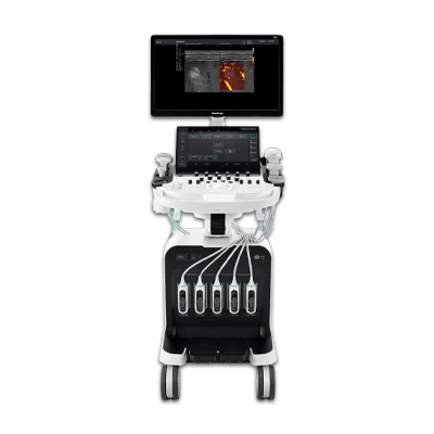
Best Prostate Ultrasound Machine in UK!
Buy and Rental Options available!
Artificial Intelligence Is Further Improving Ultrasound Accuracy
Artificial intelligence (AI) is becoming an increasingly important component of modern ultrasound systems. Rather than replacing the clinician, AI assists by analysing image patterns that may be difficult for the human eye to recognise consistently. Current AI applications include:
- Automatic prostate segmentation
- Suspicious lesion detection
- Image quality optimisation
- Needle tracking during biopsy
- Measurement automation
- Standardised reporting
Machine learning algorithms can also reduce operator variability by providing consistent image analysis across different users and clinical settings.
Researchers are actively developing AI systems capable of highlighting suspicious regions during real-time ultrasound scanning. While many of these technologies are still being validated, early results are promising and suggest AI may improve both efficiency and diagnostic consistency (European Urology Open Science; PubMed).
Operator Experience Still Matters
Even the most advanced ultrasound equipment depends on the skill of the person using it. Unlike CT or MRI, ultrasound is highly operator-dependent. The quality of the examination can vary based on:
- Probe positioning
- Image optimisation
- Understanding of anatomy
- Recognition of artefacts
- Needle alignment during biopsy
- Interpretation of abnormal findings
Experienced clinicians know how to adjust scanning parameters continuously to obtain the clearest possible image. They also recognise common artefacts that could otherwise be mistaken for pathology.
Training, standardised biopsy protocols, and the use of accessories such as stepper stabilisers and needle guides help improve consistency between operators.
Equipment Quality Also Influences Accuracy
Image quality is not determined by the ultrasound probe alone. Modern ultrasound systems incorporate numerous technologies designed to improve visualisation, including:
- Digital beamforming
- Speckle reduction filters
- Adaptive image processing
- Compound imaging
- Tissue harmonic imaging
- High-resolution monitors
Regular equipment maintenance and calibration also contribute to reliable image quality. Even small reductions in transducer performance can affect spatial resolution and lesion visibility.
Factors Reduce Ultrasound Image Accuracy
Although ultrasound is an extremely valuable imaging modality, several factors can reduce image quality. These include:
Patient Factors
- Obesity
- Excess bowel gas
- Previous pelvic surgery
- Anatomical variation
Technical Factors
- Incorrect gain settings
- Poor probe positioning
- Limited acoustic window
- Low-frequency imaging when higher frequencies are appropriate
Operator Factors
- Limited experience
- Inadequate image optimisation
- Failure to recognise artefacts
- Inaccurate needle alignment
Understanding these limitations helps clinicians adapt their technique and improve diagnostic confidence.
Conclusion
Ultrasound has evolved from a basic imaging tool into a sophisticated technology that plays a central role in modern prostate cancer diagnosis. Its ability to provide real-time visualisation, optimise images during scanning, and guide biopsy procedures has significantly improved diagnostic accuracy and procedural confidence.
Advances such as high-frequency transducers, harmonic imaging, Doppler ultrasound, elastography, contrast-enhanced ultrasound, MRI-ultrasound fusion, micro-ultrasound, and AI-assisted image analysis are transforming how clinicians detect and target clinically significant prostate cancer. While no single technology is perfect, combining high-quality ultrasound systems with experienced operators and evidence-based biopsy techniques offers the best opportunity for accurate diagnosis, fewer repeat procedures, and improved patient outcomes.
Book your appointments with our expertise for any kind of queries.
FAQs
1. Is ultrasound more accurate than MRI?
They serve different purposes. MRI generally provides superior soft tissue contrast for identifying suspicious prostate lesions, while ultrasound offers real-time guidance during biopsy. When combined through MRI-ultrasound fusion, the strengths of both technologies can significantly improve biopsy accuracy.
2. Does ultrasound improve prostate biopsy accuracy?
Yes. Ultrasound provides continuous real-time guidance, allowing clinicians to visualise the prostate and biopsy needle throughout the procedure. This improves targeting accuracy, reduces sampling errors, and increases confidence that tissue has been collected from the intended location.
3. What is micro-ultrasound?
Micro-ultrasound is a high-frequency ultrasound technology that operates at around 29 MHz. It provides substantially higher image resolution than conventional ultrasound, allowing clinicians to visualise prostate tissue in greater detail and improve lesion detection.
4. Can ultrasound detect prostate cancer on its own?
Ultrasound can identify suspicious abnormalities, but it cannot reliably diagnose prostate cancer by itself. A biopsy is required to confirm the diagnosis. Newer techniques such as micro-ultrasound, elastography, and contrast-enhanced ultrasound improve lesion detection but are still used alongside biopsy.
5. Why is ultrasound preferred during prostate biopsy?
Ultrasound provides immediate visual feedback throughout the procedure. Clinicians can monitor needle placement in real time, adjust the trajectory if necessary, and accurately target suspicious regions while avoiding surrounding structures.