Author name: Mr Bachar Zelhof

Mr. Bachar Zelhof is a Consultant Urological Surgeon at Manchester University NHS Foundation Trust and a leading Northwest expert in robotic kidney surgery, kidney stones, and complex endometriosis. An accomplished academic with over 30 publications and an MD with honours, he serves as a Senior Honorary Lecturer at the University of Manchester and was named the 2021 Northwest Urology Trainer of the Year.

brachytherapy

Functions of Brachy Grid: A Complete Guide to Its Role in Prostate Procedures

Brachy grids play a critical role in modern prostate procedures by helping clinicians place needles with exceptional accuracy during prostate brachytherapy and transperineal prostate biopsy. Used alongside an ultrasound probe and stepper stabiliser, a brachy grid provides a fixed coordinate system that improves precision, treatment consistency, and patient safety. What Is a Brachy Grid? A brachy grid, also known as a template grid or perineal template, is a medical device used to guide needle placement through the perineum during prostate procedures. It is mounted onto a stepper stabiliser and aligned with the probe, creating a precise coordinate system that allows clinicians to target specific areas of the prostate. Most brachy grids feature a matrix of evenly spaced holes, commonly 5 mm apart, although some systems use 3 mm spacing for finer targeting. Each hole is identified by a letter and number, enabling accurate documentation and repeatable needle placement. The grid is most commonly used in: According to the American Association of Physicists in Medicine (AAPM) and peer-reviewed literature, template-guided needle placement has become a standard component of modern prostate brachytherapy because it improves implant accuracy and dosimetric outcomes. What are the functions of Brachy Grid? 1. Provides Precise Needle Guidance The primary function of brachy grid is to guide needles into the prostate with a high level of precision. Rather than relying on freehand needle placement, clinicians insert the biopsy gun through a predefined hole in the grid. The grid acts as a physical guide, helping maintain the planned entry point while reducing unwanted movement. This level of precision is particularly important because even a small deviation can affect seed placement or tissue sampling. Studies published in Brachytherapy and Medical Physics have shown that template-guided procedures improve needle placement accuracy and contribute to better treatment quality. 2. Creates a Coordinate Mapping System Every hole within the grid corresponds to a specific coordinate. During ultrasound imaging, physicians identify the target location inside the prostate and match it with the appropriate grid coordinate.  This allows every needle position to be documented accurately throughout the procedure. The coordinate system also enables: This structured mapping is one of the reasons template-guided procedures have become widely adopted in prostate brachytherapy. 3. Improves Seed Placement During Brachytherapy In prostate brachytherapy, radioactive seeds or temporary radiation catheters must be placed exactly where the treatment plan specifies. The brachy grid ensures needles enter the prostate at the planned locations, helping clinicians distribute radiation evenly throughout the gland while minimising exposure to surrounding structures such as the urethra, bladder and rectum. Accurate seed placement contributes to: Research published in Radiotherapy and Oncology has consistently shown that implant accuracy is a major factor influencing successful prostate brachytherapy outcomes. 4. Supports Transperineal Prostate Biopsy Beyond brachytherapy, brachy grids are increasingly used during template-guided transperineal prostate biopsy. The grid allows systematic sampling of the prostate by guiding biopsy needles through predefined locations. This approach offers several advantages over traditional transrectal biopsy: Current European Association of Urology (EAU) guidelines recognise transperineal biopsy as an effective approach for prostate cancer diagnosis, with growing adoption worldwide. 5. Enhances Procedure Reproducibility One of the biggest advantages of a brachy grid is consistency. Every procedure follows the same coordinate system, making results reproducible regardless of operator experience. This standardisation is valuable for: Standardised needle placement also helps reduce variability between procedures. 6. Works Together with Ultrasound Imaging The brachy grid is not used independently. It functions alongside: The ultrasound provides real-time visualisation of the prostate, while the stepper maintains stable probe positioning. The brachy grid translates the ultrasound image into physical coordinates, enabling clinicians to guide needles accurately to the intended target. This integration forms the foundation of modern image-guided prostate interventions. 7. Helps Protect Surrounding Structures Precise needle placement reduces unnecessary trauma to surrounding tissues. By following planned needle paths, clinicians can better avoid important structures including: This contributes to safer procedures while supporting treatment accuracy. 8. Improves Workflow in the Operating Room A brachy grid also improves procedural efficiency. On the stepper system, the grid provides a stable reference throughout the procedure. Instead of estimating needle positions repeatedly, clinicians simply select the appropriate coordinate. The benefits include: These workflow improvements become particularly valuable during complex HDR brachytherapy procedures involving multiple needles. Why does hole spacing matter? Most prostate brachy grids use 5 mm spacing, although specialised applications may use smaller intervals. The spacing has been developed through decades of clinical experience and remains a key design feature of modern template systems. Uniform spacing allows clinicians to: Conclusion The brachy grid is an essential component of modern image-guided prostate procedures. By providing a precise coordinate system for needle placement, it improves the accuracy of brachytherapy, supports systematic transperineal biopsy, enhances procedural consistency, and helps protect surrounding healthy tissues. When combined with a stepper stabiliser, ultrasound imaging, and appropriate treatment planning, the brachy grid enables clinicians to perform prostate interventions with a high degree of precision. As prostate cancer diagnosis and treatment continue to evolve, the brachy grid remains a cornerstone of safe, reproducible, and effective prostate care. FAQs

product blog

Needle for prostate biopsy | What kind of needle is used?

If your doctor has recommended a prostate biopsy, you may wonder what kind of needle is used for a prostate biopsy. A modern transperineal prostate biopsy uses three specialised instruments: a spinal needle for local anaesthesia, a coaxial needle to create an access pathway, and a prostate biopsy gun to collect tissue samples. Learn more.

Home

Fixed-Angle vs Freehand Transperineal Biopsy: Which is More Accurate?

As transperineal prostate biopsy becomes the new standard of care across the UK and internationally, urology departments face a second decision: Fixed-Angle vs Freehand Transperineal Biopsy: Which is More Accurate? Both approaches share the same core advantage of avoiding the rectum and eliminating sepsis risk. However, they differ significantly in how the needle is guided, how reproducible the results are, and how quickly a clinical team can adopt them. For urologists, procurement heads, and department leads evaluating their transperineal pathway, understanding the difference between fixed-angle and freehand techniques is critical to achieving consistent, high-quality diagnostic outcomes. What is Freehand Transperineal Biopsy? Freehand transperineal biopsy involves the urologist manually inserting the biopsy needle through the perineum without a mechanical guide or grid to restrict the angle of entry. The clinician uses real-time ultrasound imaging to direct the needle by hand toward the target zone of the prostate. This approach offers flexibility and requires no additional guiding hardware beyond the ultrasound probe. However, it places significant demands on the operator’s skill and spatial awareness, as the needle trajectory is entirely dependent on individual technique. Key characteristics of freehand transperineal biopsy: What is Fixed-Angle Transperineal Biopsy? Fixed-angle transperineal biopsy uses a precision-engineered needle guide, such as Surefire or Innofine Needle Guide, that attaches to the ultrasound probe and locks the needle entry at a predetermined, consistent angle. The biopsy needle travels through the guide at a fixed trajectory, allowing the urologist to systematically sample all regions of the prostate with reproducible accuracy. This mechanical constraint is not a limitation; it is a clinical advantage. By removing the variable of hand angle, fixed-angle systems standardise sampling across operators, procedures, and centres. Key characteristics of fixed-angle transperineal biopsy: Clinical Accuracy: Fixed-Angle vs Freehand Transperineal Biopsy A key concern for every urology department is determining which technique is more effective at identifying clinically significant prostate cancer. Clinical Parameter Freehand Fixed Angle Anterior Zone access Variable Consistent Systematic sampling Operator dependent Standardised Cancer detection rate Lower in less experienced High and reproducible Operator variability High Minimal MRI fusion compatibility Limited High Learning curve Steep Shorter Repeat biopsy reproducibility inconsistent Consistent Fixed-angle systems provide a significant advantage in anterior prostate sampling. The anterior zone is one of the most commonly missed areas in any biopsy approach, and it is where a meaningful proportion of clinically significant cancers reside. Because the fixed-angle guide allows systematic grid-based targeting, it ensures this region is sampled reliably in every procedure. In freehand technique, anterior access depends on the operator’s ability to manually angle the needle through a narrow perineal entry point, a skill that takes considerable time to develop and is difficult to standardise across a department. Workflow and Training Considerations For department leads overseeing multiple urologists with varying levels of transperineal experience, standardisation of technique is as important as the technique itself. For NHS trusts and private hospital groups transitioning their entire urology department to transperineal biopsy, fixed-angle systems reduce the risk of inconsistent outcomes during the transition period. Fixed-Angle Needle Guide by Global Prostate Solutions At Global Prostate Solutions, the Needle Guides are engineered specifically for fixed-angle transperineal biopsy. Both devices attach to the ultrasound probe and lock the needle at a precise angle, enabling: The fixed-angle Needle Guide removes variability from the equation. Whether a procedure is performed by a consultant urologist, the guide ensures the needle follows the same precise trajectory every time. Consult the best urologist in Manchester for queries related to urology. Conclusion Both fixed-angle and freehand transperineal biopsy eliminate the core risk of the transrectal approach, rectal bacterial contamination, and post-biopsy sepsis. However, when it comes to diagnostic accuracy, departmental consistency, and ease of training, fixed-angle systems hold a clear clinical and operational advantage. For urology departments committed to delivering the highest standard of prostate cancer diagnostics across consultants, trainees, and high-volume procedures, the GPS Fixed-Angle Needle Guide provides the mechanical precision and reliability that freehand technique simply cannot guarantee. FAQs

brachytherapy

What is LDR Brachytherapy? Know it’s step by step procedure

Low-dose rate (LDR) brachytherapy is a method of delivering radiation at a rate of less than 2.0 Gy/h by placing sealed radiation sources directly in or near the treatment area. In plain terms, it is an internal form of radiation therapy; “brachy” comes from the Greek word for “short distance,” reflecting that the radiation source is placed very close to or within the tumour itself.  What is LDR Brachytherapy? LDR brachytherapy is a type of internal radiation treatment used to treat localised prostate cancer. During the procedure, tiny radioactive seeds are placed directly into the prostate gland, where they slowly release radiation over time. Since the radiation is focused directly on the cancer, it can destroy tumour cells more precisely while helping to protect nearby healthy tissues, such as the bladder and rectum.  This treatment is considered minimally invasive and is usually completed in a single procedure, allowing many patients to return to their normal routine more quickly compared to surgery or several weeks of external beam radiotherapy. LDR brachytherapy is also known for offering good long-term cancer control outcomes, while helping many men maintain better urinary continence and sexual function than some alternative treatments. Unlike prostate removal surgery, which completely removes the prostate gland, brachytherapy treats the cancer from inside the body without major incisions. It may be recommended for men with localised or low-to-intermediate risk prostate cancer, depending on their overall health and cancer stage. How does LDR Brachytherapy help treat prostate cancer? Low-dose rate (LDR) brachytherapy, often called seed implantation or prostate brachytherapy, is a form of internal radiation treatment used for prostate cancer. The procedure involves placing tiny radioactive seeds directly into the prostate gland, allowing radiation to target the cancer more precisely while limiting exposure to nearby healthy tissues. As the treatment is minimally invasive, it does not involve major surgery, and most patients only need a short hospital stay, often returning home the same day or within 24 hours. Recovery is generally quick, with many men resuming their normal daily activities within a few days. LDR brachytherapy differs from external beam radiotherapy (EBRT), where radiation is delivered from outside the body using high-energy X-rays. In EBRT, the radiation must pass through surrounding tissues before reaching the prostate, which can sometimes affect nearby healthy areas. In contrast, brachytherapy delivers radiation from inside the prostate itself, helping concentrate treatment directly on the tumour site. What is the procedure? Before undergoing LDR brachytherapy, patients usually attend a consultation with an oncologist to prepare for treatment. Depending on the hospital, this planning appointment may take place a few days or weeks before the procedure, while some centres complete the planning on the same day as treatment. At this stage, doctors may use imaging techniques like ultrasound or MRI scans. It helps in providing a more detailed view of the prostate gland. These images help the medical team identify the exact location of the tumour and calculate the most suitable radiation dose for treatment. The procedure itself is carried out under either general anaesthesia or spinal anaesthesia. Small radioactive seeds, each roughly the size of a grain of rice, are carefully placed into the prostate through fine hollow needles. Ultrasound guidance is commonly used throughout the procedure to ensure accurate placement of the seeds. The needles are inserted through the perineum, which is the area between the scrotum and the anus, and removed once the seeds are positioned correctly. LDR brachytherapy is typically completed within about an hour. After recovering from the anaesthetic, most patients can return home later the same day. A follow-up CT scan may be arranged within several weeks after treatment to review the placement of the seeds and assess the treatment plan further. The implanted seeds remain inside the prostate and gradually release radiation over several months. This targeted approach helps destroy cancer cells while reducing exposure to surrounding healthy tissues, allowing many patients to continue with their normal daily activities with minimal disruption. Who is suitable for this procedure? LDR brachytherapy is often recommended for men with localised prostate cancer, meaning the cancer is contained within the prostate gland. It may be suitable for patients with low-risk or intermediate-risk prostate cancer, depending on factors such as: In some cases, LDR brachytherapy may be combined with other treatments, including hormone therapy or external beam radiotherapy. A specialist will assess whether the treatment is appropriate based on each patient’s individual condition. What are the benefits? LDR brachytherapy offers several advantages for suitable patients, including: The procedure does not involve major surgical incisions, which can reduce recovery time and hospital stay. Radiation is delivered directly into the prostate, helping reduce exposure to nearby healthy organs and tissues. Most patients return to normal daily activities within a few days following treatment. Many men are discharged on the same day as the procedure or after a short overnight stay. LDR brachytherapy has demonstrated strong long-term outcomes in appropriately selected patients with localised prostate cancer. Compared with some other prostate cancer treatments, many patients experience lower rates of urinary incontinence and preservation of sexual function. What are the possible side effects of LDR Brachytherapy Although LDR brachytherapy is generally well tolerated, some side effects may occur during recovery. These can include: These symptoms are usually temporary and often settle down as time goes on. However, patients should contact their healthcare team if symptoms become severe or persistent. LDR Brachytherapy vs External Beam Radiotherapy (EBRT) Both LDR brachytherapy and external beam radiotherapy are used to treat prostate cancer, but they deliver radiation differently. In external beam radiotherapy, radiation is directed at the prostate from outside the body using high-energy X-rays. The radiation must pass through surrounding tissues before reaching the tumour. In contrast, LDR brachytherapy places radioactive seeds directly inside the prostate. This allows radiation to target the tumour more precisely while limiting exposure to nearby healthy structures. Another difference is treatment duration. External beam radiotherapy often requires multiple hospital visits over several weeks, while LDR brachytherapy is usually completed

Home, Prostate Health

Prostate Biopsy Under Local Anaesthesia| How to perform

A prostate biopsy is one of the most important procedures used to diagnose prostate cancer and investigate abnormal prostate findings. Traditionally, many prostate biopsies were performed under sedation or general anaesthesia, but advances in medical techniques now allow many men to safely undergo a prostate biopsy under local anaesthesia in an outpatient setting. This modern approach is minimally invasive, well tolerated by most patients, and allows for quicker recovery while maintaining excellent diagnostic accuracy. In particular, transperineal prostate biopsy under local anaesthesia is increasingly being adopted worldwide because of its lower infection risk and improved patient safety.  What is a Prostate Biopsy? A prostate biopsy is a procedure in which small tissue samples are taken from the prostate gland and examined under a microscope to check for cancer or other abnormalities. Doctors may recommend a prostate biopsy if: What is Prostate Biopsy under Local Anaesthesia? Local anesthesia means the area being treated is numbed while the patient remains awake during the procedure. Unlike general anesthesia, patients do not need to be fully unconscious. During a prostate biopsy, the doctor injects local anesthetic medication into the biopsy area to reduce discomfort. Patients may still feel pressure or mild sensations, but significant pain is usually minimised. Many modern transperineal prostate biopsies can now be safely performed under local anesthesia in a clinic or outpatient setting.  Why is local anesthesia becoming more popular? Local anesthesia offers several benefits compared to general anesthesia or sedation. How is a Prostate Biopsy under Local Anesthesia performed? Although techniques may vary slightly between hospitals and clinics, the procedure generally follows these steps: Before the biopsy: The patient lies comfortably while the doctor prepares the biopsy area. The skin and deeper tissues are numbed using local anesthetic injections. Patients may feel a brief stinging sensation during this step. An ultrasound probe is inserted into the rectum to guide accurate needle placement. Small biopsy samples are taken from targeted areas of the prostate. MRI-targeted biopsies may also be performed if suspicious lesions are identified on imaging. The entire procedure usually takes around 20–45 minutes, depending on the number of samples required.  Does a local anesthesia for prostate biopsy hurts? Most men experience mild to moderate discomfort rather than severe pain. The anesthetic injections are often the most uncomfortable part of the procedure. Once the area becomes numb, many patients report only pressure sensations or brief discomfort during sampling. Patient experiences can vary depending on anxiety levels, prostate size, biopsy technique, and individual pain tolerance. Community experiences shared online also suggest that while some men find the procedure uncomfortable, many tolerate it better than expected under local anesthesia.  Benefits of Transperineal Prostate Biopsy Under Local Anesthesia What are the possible side effects and risks? Although prostate biopsy is generally safe, some temporary side effects are common. Common Side Effects: These symptoms usually improve within days to weeks.  Less Common Risks: Patients should contact their doctor immediately if they develop fever, severe pain, inability to urinate, or heavy bleeding after the procedure. Recovery after a Prostate Biopsy Recovery after a local anesthetic biopsy is usually straightforward. Most patients are advised to: Blood in semen can sometimes persist for several weeks and is usually not dangerous. Who may benefit most from this procedure? Transperineal prostate biopsy under local anesthesia may be especially helpful for: Is Local Anesthetic Prostate Biopsy effective? Yes, Studies show that transperineal biopsy under local anesthesia provides excellent cancer detection rates while improving patient safety and reducing the risk of infection. Many specialists now consider it one of the preferred modern biopsy approaches.  Conclusion Prostate biopsy under Local Anesthesia has become a safe, effective, and minimally invasive option for diagnosing prostate cancer. With lower infection risk, shorter recovery time, and improved patient comfort, transperineal biopsy under local anesthesia is increasingly being preferred by both patients and specialists. Although mild discomfort may occur, modern techniques have made the procedure more accurate and better tolerated than ever before. Consult the best urologist in Manchester to know more about the prostate biopsy procedure. FAQs

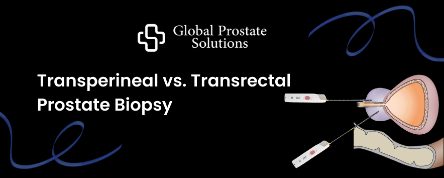
Transperineal vs. Transrectal Prostate Biopsy
Case Studies

Why are urologists shifting from Transrectal to Transperineal Prostate Biopsy?

For decades, the transrectal biopsy has been the standard method for diagnosing prostate cancer. However there were increased concerns regarding infection risk, antibiotic resistance, and diagnostic limitations. Healthcare providers are prioritising safer, more accurate, and cost-effective approaches in their clinical settings. In the field of urological diagnostics, the shift from Transrectal Ultrasound-guided (TRUS) biopsies to Transperineal (TP) biopsies represents one of the most significant safety and accuracy upgrades in recent clinical history. For procurement heads and department leads, the transition is no longer just a clinical preference—it is a strategic move to reduce post-operative complications and long-term hospital costs. What is Transrectal Prostate Biopsy? A transrectal prostate biopsy is a procedure in which a needle is passed through the wall of the rectum into the prostate to collect tissue samples, usually guided by ultrasound. Since the needle passes through the rectum, there is a higher risk of infection, and antibiotics are typically required. Challenges with Transrectal Biopsy The transrectal approach involves passing the biopsy needle through the rectal wall, which introduces bacteria into the prostate and bloodstream. This significantly increases the risk of infection and necessitates the routine use of prophylactic antibiotics, contributing to growing global concerns around antibiotic resistance. This method is associated with a higher rate of post-procedure complications. Parameter Data Infection / Sepsis Rate 2% – 7% Hospital Admissions 1% – 3% Antibiotic Usage 100% of patients Risk Factor Bacterial contamination via rectum What is Transperineal Prostate Biopsy? A transperineal prostate biopsy involves inserting the needle through the skin between the anus and scrotum (the perineum) to reach the prostate. This approach avoids the rectum, significantly reducing the risk of infection and improving access to all areas of the prostate. It is increasingly preferred by 50% of urologists across the UK due to its safety, better diagnostic accuracy, and reduced need for antibiotics. Advantages of Transperineal Biopsy The transperineal approach eliminates rectal involvement by accessing the prostate through the perineal skin, significantly improving safety and clinical outcomes. (i) Near-Zero Infection Risk By avoiding the rectal pathway, the transperineal method reduces bacterial exposure, resulting in extremely low infection rates. Parameter Data Infection / Sepsis Rate <0.1% – 0.5% Clinical Outcome Many centres report zero sepsis cases (ii) Improved Diagnostic Accuracy Transperineal biopsy provides better access to the anterior region of the prostate, which is often missed in transrectal procedures. This leads to improved detection of clinically significant cancers. Method Detection Rate Transperineal ~45% – 55% Transrectal ~35% – 45% (iii) Reduced Antibiotic Dependency With minimal infection risk, transperineal biopsy can often be performed without antibiotics, addressing concerns around antimicrobial resistance. (iv) Cost Efficiency for Healthcare Systems Although initial setup costs for transperineal biopsy may be slightly higher, the reduction in complications leads to significant long-term savings. Cost Factor Transrectal Transperineal Infection-related hospitalisation £1,500 – £3,000 per case Negligible Annual financial impact High due to complications Significantly reduced Estimated annual savings after switching procedure The estimated annual savings when switching from transrectal to transperineal prostate biopsy primarily come from the significant reduction in infection-related complications. While transperineal biopsy may have a slightly higher upfront procedure cost, it dramatically lowers infection rates from around 2–7% in transrectal procedures to less than 0.5% in transperineal approaches.  This reduction leads to fewer hospital admissions, decreased antibiotic use, and minimal post-procedure complications. In a typical hospital performing around 1,000 biopsies per year, transrectal biopsy can result in approximately 30 infection cases annually, each costing between £1,500 and £3,000 for treatment. The transperineal biopsy reduces this to 0–5 cases per year, resulting in substantial cost savings. Lets understand with the help of a case example; During the Transperineal Approach After Transperineal Implementation Real World Adoption Trends Healthcare systems, particularly in the United Kingdom are rapidly transitioning toward transperineal biopsy as the new standard of care. Procurement & Clinical Benefits The transperineal prostate biopsy offers significant clinical, financial, and operational advantages. It is a much safer procedure with minimal infection risk and provides a higher detection rate of clinically significant cancers. The procedure can be performed under local anaesthesia, enables faster patient recovery, and improves overall patient throughput and workflow efficiency within healthcare settings. Conclusion The transition from transrectal to transperineal prostate biopsy represents a critical advancement in prostate cancer diagnostics. With substantially lower infection rates, improved diagnostic accuracy, and significant cost savings, transperineal biopsy is emerging as the preferred standard for modern healthcare systems. For hospitals and procurement leaders, adopting this approach is not only a clinical decision but also a strategic investment in patient safety, operational efficiency, and long-term sustainability.

Scroll to Top